Epidermoid Cyst Floor Of Mouth Radiology
Floor Of The Mouth Cystic Lesion Radiology Case Radiopaedia Org
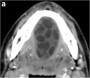
Floor Of Mouth Dermoid Cyst Radiology Case Radiopaedia Org
Floor Of Mouth Dermoid Cyst Radiology Case Radiopaedia Org

Pin On Ct

Pin By Liz On Neuro Medical Knowledge Radiology Medicine Studies
Http Pdf Posterng Netkey At Download Index Php Module Get Pdf By Id Poster Id 101707
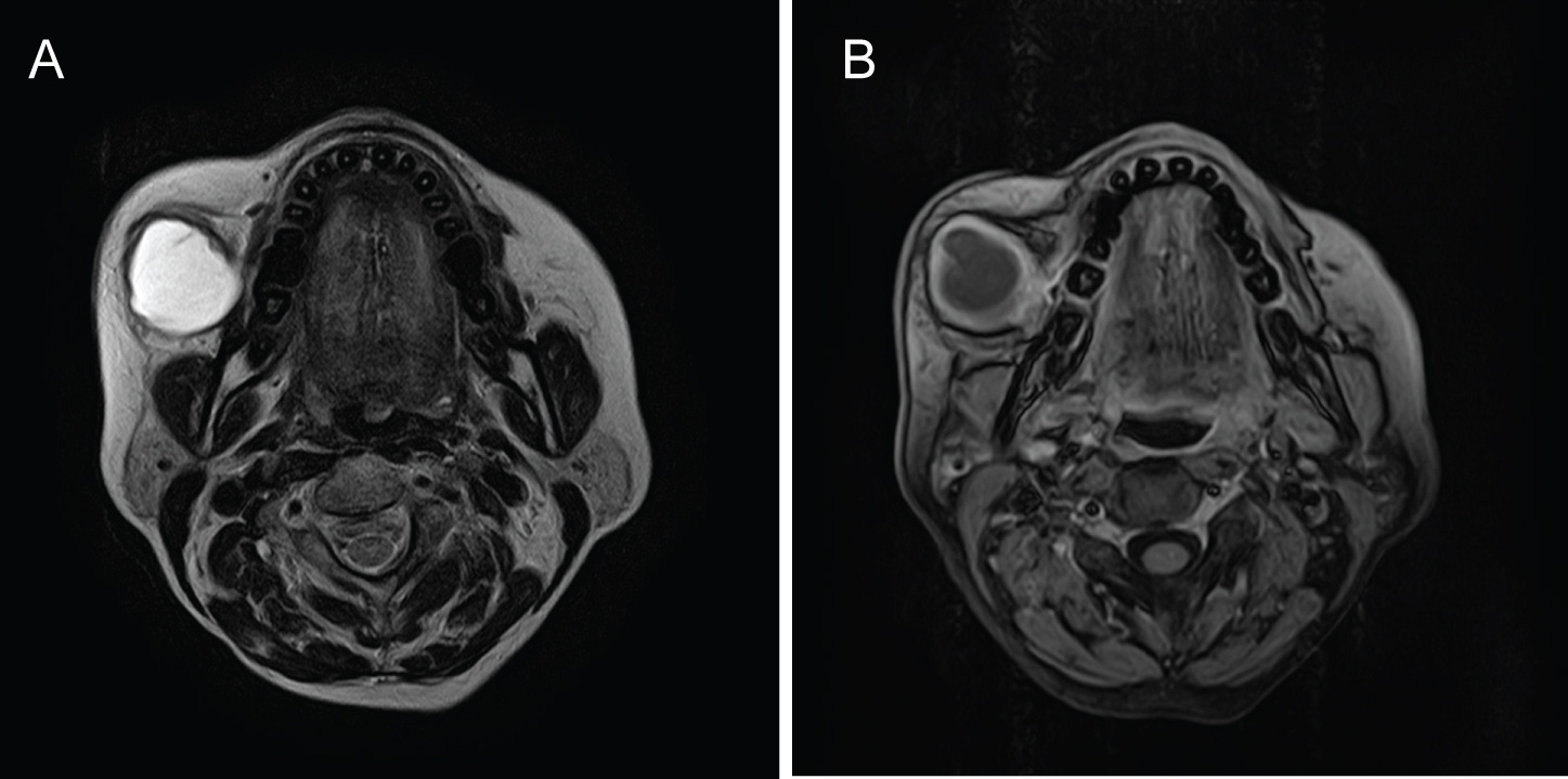
Dermoid cysts contain skin appendages and cystic teratomas present tissue originating from all the germinal layers.
Epidermoid cyst floor of mouth radiology.

Imaging The Floor Of The Mouth And The Sublingual Space Radiographics
Dermoid Cyst Mri Online

View Image

The Radiology Assistant Brain Tumor Systematic Approach Brain Tumor Tumor Brain Tissue
Http Pdf Posterng Netkey At Download Index Php Module Get Pdf By Id Poster Id 40540
Imaging Of The Sublingual And Submandibular Spaces Springerlink

Quiste Epidermoide Quiste Epidermoide
Https Pubs Rsna Org Doi Pdf 10 1148 Rg 317095738

Ecr 2013 C 1867 High Resolution Ultrasound In The Assessment Of Soft Tissue Tumors And Tumor Like Lesions Ultrasound Epidermoid Cyst Subcutaneous Tissue

Epos

Pdf Duck Egg Size Epidermoid Cyst In The Floor Of Mouth A Rare Entity Semantic Scholar

The Lesion Is Most Compatible With A White Epidermoid Cyst Epidermoid Cysts Are Typically T1 Isointense And Flair Hypointense Epidermoid Cyst Cysts Radiology

Figure 5 From Case Report Of Complicated Epidermoid Cyst Of The Floor Of The Mouth Radiology Histopathology Correlation Semantic Scholar

Dermoid Cyst Of The Parotid Gland Report Of A Rare Entity With Radiological Findings And Treatment Approaches

Pin By Dr Abuaiad On Brain Head And Neck With Images Molar Tooth Radiology Head And Neck
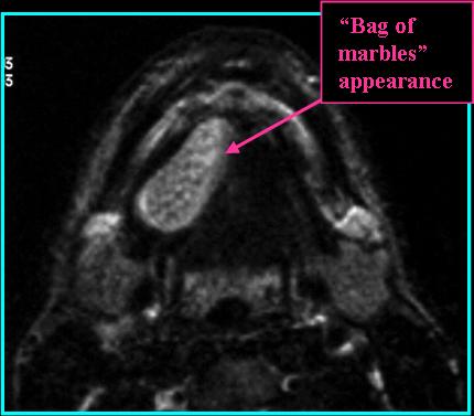
Dermoid Cysts Rads Iowa Head And Neck Protocols

Cavum Velum Interpositum Cyst Radiology Case Radiopaedia Org

Ranula Radiology Reference Article Radiopaedia Org
Https Encrypted Tbn0 Gstatic Com Images Q Tbn 3aand9gctl3q2ghmbp9p6sit2xyysmhj6o Ojxcxuuutxxr9s Nd2xi3rm Usqp Cau
Laryngeal Cyst Radiology Reference Article Radiopaedia Org

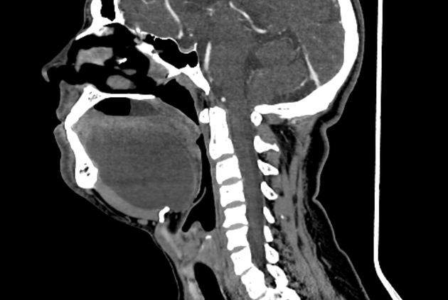
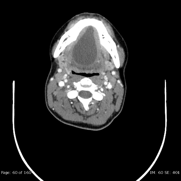
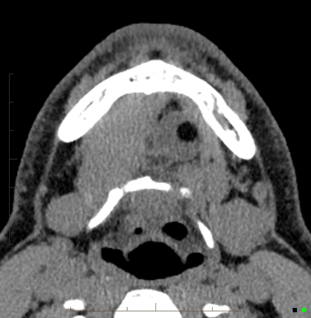
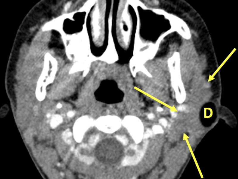
Contrast Ct In A Teenager With A Palpable Mass In The Right Side Of The Mouth For About A Year Shows A Unilocular Fluid Attenuation Mass In The Right Sublingual Con Imagenes

Dermoid Cysts Once Upon A Time Academic Dermatology Of Nevada

Cavernous Hemangioma Cavernoma Radiology Radiology Imaging Neurology

Primary Lesions Of The Root Of The Tongue Radiographics
Source : pinterest.com
